Region detection
Coarse analysis proposes tumour-bearing regions on the whole-slide image, giving the pathologist a starting point for closer inspection.

AI-assisted digital pathology. Expert judgment at the centre.
PathAI connects whole-slide imaging, three AI capabilities and pathologist-led review for colorectal adenocarcinoma, from case intake to a finalized report.
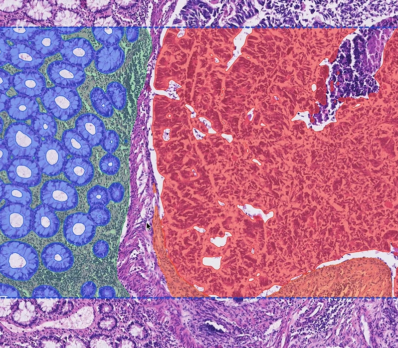
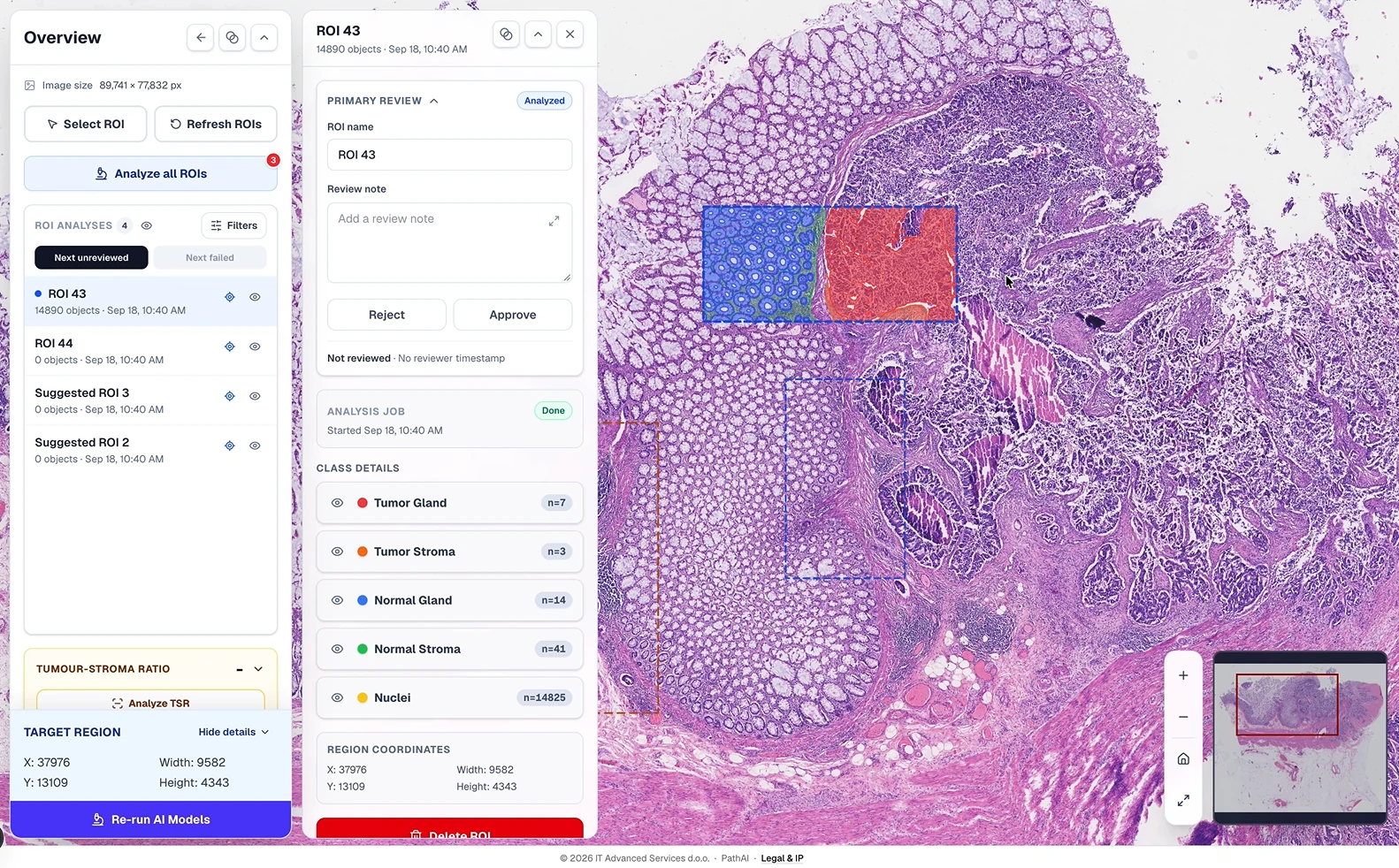
Draw a region, then run the demo to reveal four tissue classes.
Synthetic slide · precomputed result. This promotional demo uses predefined overlays, without live AI inference or uploaded slides. The full platform’s nucleus analysis, tumour–stroma ratio and expert approval workflow are outside this demonstration.
Coarse analysis proposes tumour-bearing regions on the whole-slide image, giving the pathologist a starting point for closer inspection.
Detailed analysis distinguishes tumour glands, tumour stroma, normal glands and normal stroma, with background handled separately.
Individual nuclei are identified and measured, producing counts, area, shape and density information alongside tissue-level findings.
Organize patient and case information, upload whole-slide images and resume interrupted uploads.
Process suggested or manually selected regions, with visible job progress and separate tissue and nucleus results.
Compare overlays with the original slide. Edit annotations, add notes, and approve or reject findings.
Request a second opinion when needed, then finalize the professional report and retain the case history.
PathAI brings tissue areas, object counts and nucleus morphology into region and case summaries. The tumour–stroma ratio is calculated from segmented tumour-gland and tumour-stroma areas.
Measurements retain analysis coverage and model provenance, so the pathologist can assess both the result and the tissue it represents. These outputs support expert interpretation within the review workflow.
Access follows institutional membership, role and case assignment. An invited external reviewer can provide a second opinion, with their annotations retained separately from the primary record.
Persistent analysis jobs support progress tracking and recovery. Versioned models, annotation revisions and audit history connect results to the processing and review actions behind them.
PathAI is an integrated research prototype developed by ITAS with the Clinical Center of Montenegro. Technical verification was followed by a final operational evaluation on 31 August 2026, documented in a joint assessment signed by three participating pathologists.
The positive assessment highlighted tumour-region detection, quantitative analysis including tumour–stroma ratio, and the ability to inspect and correct AI findings. Experts recommended further refinement in heterogeneous tumour regions and validation on a larger, more diverse case set.
Broader clinical validation, prospective studies of diagnostic performance and time savings, and further institutional integration are next steps before wider routine diagnostic use.
IT Advanced Services d.o.o.

Clinical Center of Montenegro

Innovation Fund of Montenegro
AI System for Pathological Analysis of Adenocarcinoma (PathAI) · CGI-017-24
Talk to us about applying AI to your field.