
Digital pathology opens up new ways to inspect tissue images and turn complex visual information into structured measurements. At ITAS, our work with the Clinical Center of Montenegro explores how AI can support this process while keeping pathologists in control.
AI as an ally in pathology
PathAI combines tumour-region proposals, tissue segmentation and nucleus analysis in an interactive whole-slide viewer. These tools help experts review AI findings alongside the original image and correct outputs where needed.
Capabilities and intended benefits
- Focused review — region proposals direct attention to areas for closer inspection.
- Quantitative information — tissue and nucleus analysis provide measurements, including tumour–stroma ratio, for expert interpretation.
- A connected workflow — case handling, review, corrections and reporting bring the analysis into a single application.
These capabilities are intended to support more consistent and efficient work. Faster diagnosis, fewer diagnostic errors and improved patient outcomes have not been established by the PathAI operational evaluation and require separate clinical studies.
From research to further validation
The integrated research prototype received a positive operational assessment at KCCG on 31 August 2026, signed jointly by three pathologists. Broader clinical validation, prospective workflow studies and further institutional integration remain next steps before wider routine diagnostic use.
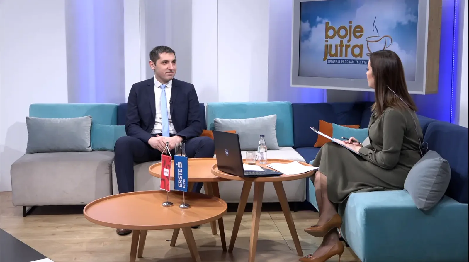
Our CEO Ivan Bošković discussed the role of AI in modern pathology on TV Vijesti. The focus of ITAS’s work is practical collaboration between software engineers and domain experts, with evidence guiding each next stage of development.